


2025
01.02
Introduction
Vesical calculus in tortoises is a significant health issue that can arise due to various factors, primarily dehydration and dietary imbalances. These calculi are primarily composed of uric acid and can lead to serious complications if not treated appropriately. If left untreated, they can cause obstruction of the cloaca, leading to difficulties in urination, constipation, pain, and even kidney damage. Symptoms of vesical calculus in tortoises include straining during urination, reluctance to urinate, and in severe cases, the presence of blood in the urine. Proper hydration, a balanced diet with adequate calcium and other essential nutrients, and prompt veterinary intervention are crucial in managing and preventing this condition.
Case 1
This is a 3-year-old Sulcata tortoise weighing 770 grams. The chief complaint is cloaca swollen and there seems to be an obstruction that cannot be passed today. The animal exhibits decreased mental alertness, reduced appetite, and decreased activity.
 |
 |
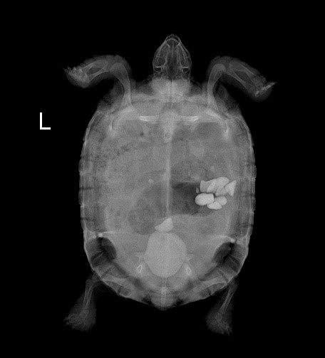
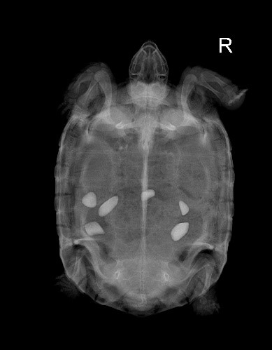
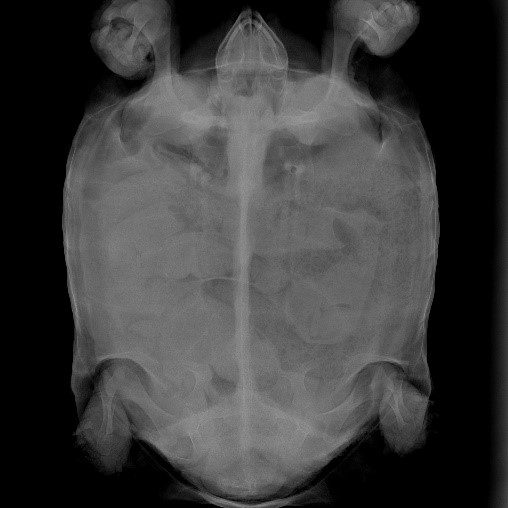
| 》Figure 1: The X-ray reveals one area of calcification. | 》Figure 2: Obstructing stones can be seen in the cloaca. |
Preoperative hematological tests show that the animal's uric acid level is elevated above the normal range, likely due to pain or discomfort caused by the obstruction, leading to a reluctance to eat or drink, resulting in dehydration. The elevated AST level may be due to smooth muscle injury caused by gastrointestinal foreign bodies and muscle damage resulting from cloacal obstruction.
》Table 1: Case 1 Biochemistry results
| Test Item | 7/21 | 7/4 | REF. Range/units |
| PCV (%) | 21 | 24 | 9 - 43% |
| GLU | 148 | 183 | 38 - 277 mg/dL |
| AMY | 2140 | 2230 | 399 - 2240 U/L |
| TBA | 1 | 1 | 0 -5.4 μmol/L |
| PHOS | 1.8 | 1.8 | 1.1 - 9.4 mg/dL |
| UA | 0.9 | > 30.0 | 0.1 - 10.5 mg/dL |
| CA | 13.3 | 13.4 | 6.5 - 20 mg/dL |
| TP | 5.3 | 5.1 | 1.5 - 7.4 g/dL |
| ALB | 2.7 | 2.8 | 0.5 - 3 g/dL |
| GLOB | 2.6 | 2.3 | 0.6 - 4.6 g/dL |
| AST | 148 | 324 | 5 - 152 U/L |
| GGT | 3 | 7 | 0 - 19 U/L |
| CHOL | 78 | 113 | 20 - 394 mg/dL |
| CK | 594 | 549 | 50 - 704 U/L |
| LDH | 593 | 685 | 100 - 977 U/L |
Postoperative care for the animal included intraperitoneal fluids, NSAID injectable medication, and laser therapy for 3 days. On the second day after treatment, the animal began eating independently but has not defecated. The animal was discharged on the third day of treatment, but the animal will continue to receive intraperitoneal fluids every two days. On the second day after discharge, the animal was observed to defecate, and the feces contained stones.
Two weeks after discharge, a follow-up hematological examination showed that all values had returned to the normal range.
 |
 |
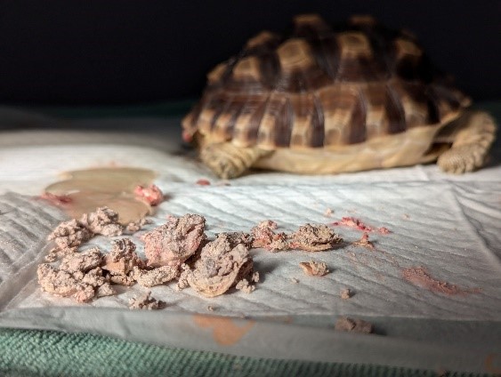
| 》Figure 3: The post-operative image shows a large amount of stones being removed. | 》Figure 4: Two weeks post-surgery, a reduction and movement of the gastrointestinal foreign body can be observed. |
Case 2
This is a 2-year-and-3-month-old Sulcata tortoise weighing 8.9 kg. The chief complaint is decreased activity for 2 days and protrusion of the penis. During the examination, a large calculi was found obstructing the cloaca, causing prolapse of the penis, which could not be retracted back into the cloaca. The penis is congested, but there is no edema on the surface, though there are slight abrasions.
 |
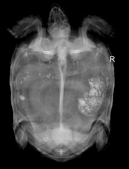
》Figure 5: The X-ray reveals one areas of calcification. A large amount of fecal material is visible in the gastrointestinal tract. |
| Preoperative blood tests revealed elevated uric acid levels, likely associated with dehydration due to decreased food and water intake. Postoperatively, the animal received intraperitoneal fluid therapy, NSAID injections, and laser treatment. After 6 days of continuous therapy, the animal was discharged. According to the owner's report, the animal's mental alertness and appetite improved after discharge, and activity levels also increased. Feces were observed for the first time 3 days after discharge. A follow-up hematological examination 14 days postoperatively showed that the uric acid levels had returned to the normal range. |
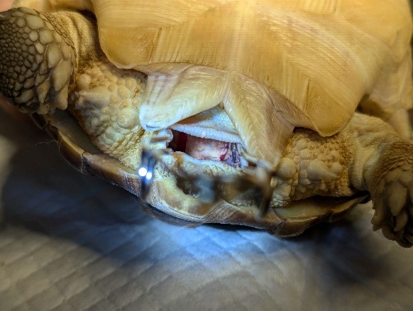
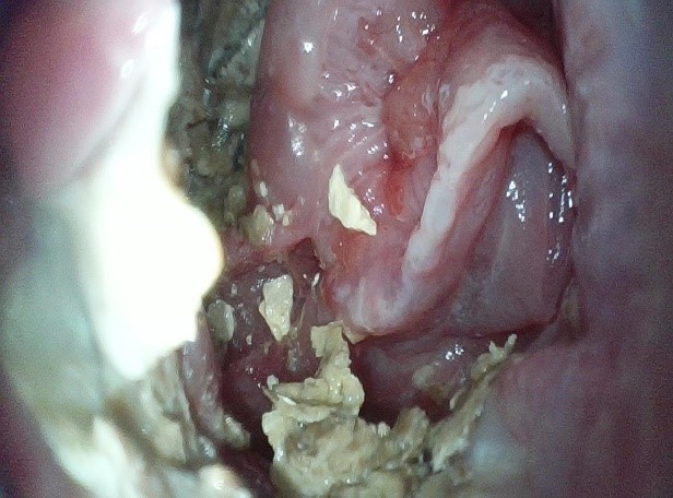
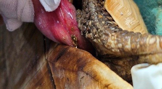 》Figure 6: The penis is congested, but there is no edema on the surface, though there are slight abrasions. |
》Table 2: Case 2 Biochemistry results
| Test Item | 9/22 | 9/8 | REF. Range/units |
| PCV (%) | 24 | 29 | 9 - 43% |
| GLU | 200 | 240 | 38 - 277 mg/dL |
| AMY | 2100 | 2103 | 399 - 2240 U/L |
| TBA | 1 | 1 | 0 -5.4 μmol/L |
| PHOS | 1.4 | 1.6 | 1.1 - 9.4 mg/dL |
| UA | 7 | 11.2 | 0.1 - 10.5 mg/dL |
| CA | 11 | 12.9 | 6.5 - 20 mg/dL |
| TP | 6 | 6.5 | 1.5 - 7.4 g/dL |
| ALB | 3.0 | 3.0 | 0.5 - 3 g/dL |
| GLOB | 3.0 | 3.5 | 0.6 - 4.6 g/dL |
| AST | 59 | 55 | 5 - 152 U/L |
| GGT | 5 | 3 | 0 - 19 U/L |
| CHOL | 20 | 20 | 20 - 394 mg/dL |
| CK | 267 | 258 | 50 - 704 U/L |
| LDH | 225 | 222 | 100 - 977 U/L |
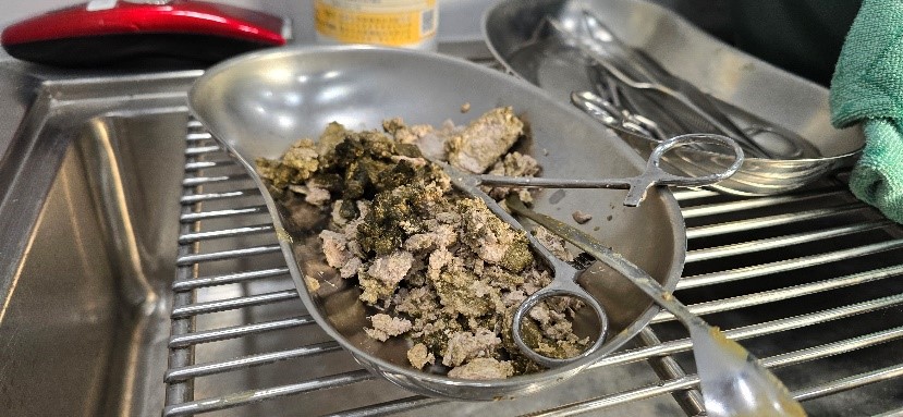 》Figure 7: A large number of calculi were removed during the surgery. |
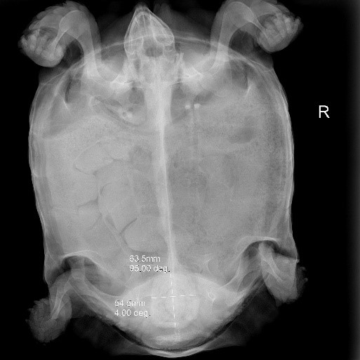
 》Figure 8: Postoperative X-ray. |
Case 3
This is an outdoor-housed Sulcata tortoise, fed leafy greens daily. Another tortoise is also kept in the household. The chief complaint is a recent decrease in the animal's activity and mental alertness. There is no heating provided outdoors. The time of the last defecation and urination is uncertain.
 》Figure 9: Multiple areas of calcification are visible on the X-ray. |
 》Figure 10: Obstructing calculi are visible in the cloaca. |
Preoperative blood tests showed elevated uric acid levels, likely related to dehydration due to reduced food and water intake. Postoperatively, the animal received intraperitoneal fluid therapy, NSAID injections, and laser treatment.
》Table 3: Case 3 Biochemistry results
| Test Item | 10/16 | REF. Range/units |
| PCV (%) | 31 | 9 - 43% |
| GLU | 182 | 38 - 277 mg/dL |
| AMY | 450 | 399 - 2240 U/L |
| TBA | 1 | 0 -5.4 μmol/L |
| PHOS | 6.4 | 1.1 - 9.4 mg/dL |
| UA | 16.3 | 0.1 - 10.5 mg/dL |
| CA | 12.0 | 6.5 - 20 mg/dL |
| TP | 6.6 | 1.5 - 7.4 g/dL |
| ALB | 2.9 | 0.5 - 3 g/dL |
| GLOB | 3.7 | 0.6 - 4.6 g/dL |
| AST | 50 | 5 - 152 U/L |
| GGT | 3 | 0 - 19 U/L |
| CHOL | 190 | 20 - 394 mg/dL |
| CK | 636 | 50 - 704 U/L |
| LDH | 674 | 100 - 977 U/L |
 》Figure 11: Multiple areas of localized erythema and edema are visible on the mucosal surface of the cloaca. |
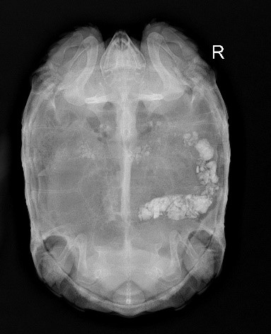 》Figure 12: Postoperative X-ray. A large amount of calcific material is visible in the gastrointestinal tract, likely representing ingested stone foreign bodies. |
On the third postoperative day, the animal was observed to be eating independently, with a noticeable improvement in its mental status, though no defecation was observed. Cucumbers and pumpkin, among other mildly laxative foods, were provided in an attempt to aid in bowel movement. The animal's activity level showed significant improvement postoperatively. The animal was discharged on the fifth postoperative day. Seven days after discharge, the owner reported that the animal had defecated and that its mental status and appetite were both normal. Although a follow-up hematological examination was initially planned for one week later, the owner indicated that financial constraints prevented further monitoring of the animal's condition.
Conclusion
Tortoises often present with nonspecific clinical symptoms, and issues are typically identified only through diagnostic examinations. Sulcata tortoises are prone to bladder calculi, which not only affect the animal's overall mental status and appetite, but can also obstruct the cloaca, leading to pain, difficulty in defecation and urination, and potentially causing mucosal ulceration. All three cases described involved calculi obstructing the cloaca, resulting in discomfort and prompting veterinary care. Owners should remain vigilant regarding their animals' feeding habits, defecation frequency, and hydration status.
References:
This case study was conducted by Chung-Jui Chen, Director of Jungle Exotic Animal Hospital, and Yong-Chen Zheng, DVM. The biochemical profile tests in cases here were measured by AmiShield veterinary chemistry analyzer and the exclusive Avian/Reptile Profile Panels.
1. Carpenter, J. W., & Harms, C. (2022). Carpenter's exotic animal formulary (6th ed.). Elsevier.
2. Nevarez, J. G. (Ed.). (2018). Blackwell's five-minute veterinary consult: Reptile and amphibian. Wiley.
3. Mader, D. R. (Ed.). (2006). Reptile medicine and surgery (2nd ed.). Saunders Elsevier.
4. Mader, D. R., & Divers, S. J. (2014). Current therapy in reptile medicine & surgery. Elsevier.
5. Chitty, J., & Raftery, A. (2013). Essentials of tortoise medicine and surgery. Wiley-Blackwell.