


2024
12.04
Introduction
The increasing popularity of pet birds, encompassing a diverse range of species from macaws to finches, has resulted in a surge of avian presentations at veterinary clinics. However, a concerning number of these cases involve malnutrition and systemic diseases, suggesting a potential knowledge gap among owners regarding proper avian care. While physical examinations and radiography are valuable diagnostic tools, these presentations often necessitate the additional sensitivity of bloodwork. Subtle clinical signs may be easily neglected, but bloodwork can reveal the mildest abnormalities, potentially leading to earlier diagnoses and improved treatment outcomes for these delicate feathered companions.
  |
Case 1
A 2-year-old emaciated female cockatiel presented for a two- to three-month history of anorexia and lethargy. The owner reported the bird experiencing a loss of appetite and decreased activity level. On presentation, the bird was observed with blepharospasm (eyes closed) and a subdued demeanor. Its plumage exhibited feather wear and darkening, and a body condition score (BCS) of 2/5, indicative of significant muscle wasting due to long-term anorexia. Labored breathing (dyspnea) suggested potential respiratory compromise.
Examination of saliva revealed no signs of upper respiratory or oral cavity infection. However, stool analysis identified light green, loose droppings, and subsequent examination of a fecal smear confirmed mild dysbiosis and poor digestion. These clinical signs, particularly the prolonged anorexia and abnormal droppings, suggested potential liver function overload. While anorexia itself could be a contributing factor, the possibility of underlying nutritional deficiencies caused by inappropriate feeding practices could not be ruled out. Further diagnostics were necessary to determine the root cause of the anorexia and the extent of liver involvement.
Radiographic examination, performed under isoflurane sedation, revealed hepatomegaly (enlarged liver) and distension of the gastrointestinal (GI) tract, suggestive of significant coelomic space occupation and potential air sac compression. Additionally, radiopaque material, consistent with mineral deposition, was visualized within the proventriculus (muscular stomach). The kidneys appeared enlarged with increased opacity, suggesting possible nephromegaly (enlarged kidneys) and potential inflammatory involvement. The enlarged liver could be attributed to fat infiltration and inflammation, often associated with an inappropriate diet lacking essential vitamins (Samiran, 2017; Dorianne, 2014).
| Following radiographic examination, blood was collected while the bird remained under anesthesia. Hematocrit (HCT) testing on a centrifuged capillary tube sample revealed a mild dehydration status (49% HCT). This dehydration likely contributed to the observed hyperalbuminemia (elevated blood albumin), while the prolonged anorexia explained the low blood glucose level. Elevated serum uric acid concentration indicated a significant decrease in glomerular filtration rate (GFR) (Harr, 2002), suggesting potential renal insufficiency and subsequent accumulation of metabolic waste products. |
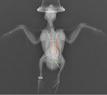
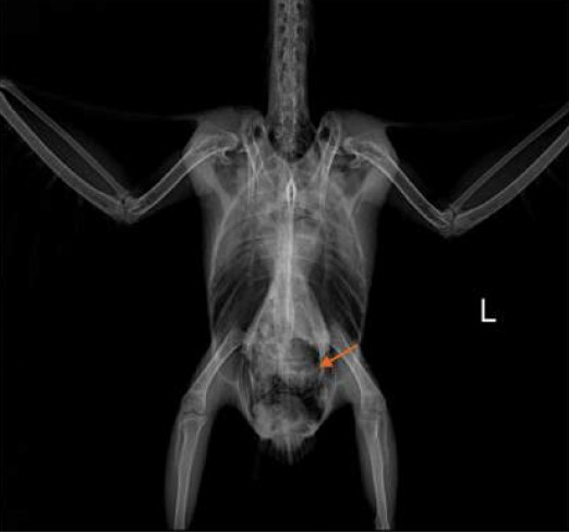
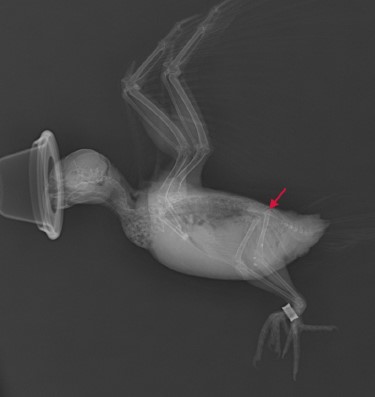 》Figure 2: Rt lateral view. Noted that the opacity and the size of the kidney both increased (red arrow)
|
》Table 1: 2024/02/01 Biochemistry results
| Test item | Result | REF. Range/units |
| Glucose | 233 | 249 - 363 mg/dL |
| TBA | 1 | 44 - 108 μmol/L |
| UA | 11.7 | 3.5 - 11 mg/dL |
| PHOS | 11.2 | 3.2 - 4.8 mg/dL |
| CA | 10.1 | 7.3 - 10.7 mg/dL |
| TP | 2.6 | 2.4 - 4.8 g/dL |
| ALB | 2.3 | 1 - 1.8 g/dL |
| GLOB | 0.3 | 0 - 9999 g/dL |
| AST | 133 | 160 - 383 U/L |
| GGT | 1 | 1 - 30 U/L |
| CHOL | 465 | 140 - 360 mg/dL |
| CK | 346 | 58 - 245 U/L |
| LDH | 50 | 120 - 455 U/L |
Analysis of liver function included measurement of total bile acids, aspartate aminotransferase (AST), and lactate dehydrogenase (LDH) activities. Both enzyme activities and total bile acid levels were markedly lower than reference ranges, which is atypical for liver damage. However, elevated serum cholesterol supported the suspicion of hepatic lipidosis (fatty liver disease). This, combined with the radiographic observation of hepatomegaly, suggests possible severe and acute liver dysfunction (Bavelaar, 2003).
Following initial diagnostics, the cockatiel was hospitalized for intensive care. The treatment plan consisted of oral medications, subcutaneous (SC) administration of lactated Ringer's (LR) fluids for hydration, and tube feeding to address the nutritional deficiencies. The medication regimen included lovastatin, a cholesterol-lowering drug, aimed at reducing hepatic lipidosis (fatty liver disease). Additionally, antioxidants were incorporated to potentially support the liver and mitigate oxidative stress.
| One week after hospitalization, a follow-up serum biochemistry profile was performed. The bird was again anesthetized with isoflurane for blood collection. The results revealed significant improvements in most parameters. Total bile acid and AST levels normalized, indicating improvement in overall liver function. Although lactate dehydrogenase (LDH) remained elevated, the increase suggested potential regeneration of liver tissue. Serum uric acid levels normalized, signifying restoration of glomerular filtration rate (GFR) and improved kidney function. Notably, serum cholesterol levels started to decline, suggesting a positive response to treatment. While a rapid return to normal cholesterol levels was not the primary goal, the treatment aimed to facilitate the body's natural processes and restore a healthy metabolic rate. |  |
》Table 2: 2024/02/09 Biochemistry results
| Test item | Result | REF. Range/units |
| Glucose | 237 | 249 - 363 mg/dL |
| TBA | 74 | 44 - 108 μmol/L |
| UA | 9.8 | 3.5 - 11 mg/dL |
| PHOS | 3.5 | 3.2 - 4.8 mg/dL |
| CA | 9.7 | 7.3 - 10.7 mg/dL |
| TP | 3.1 | 2.4 - 4.8 g/dL |
| ALB | 2.2 | 1 - 1.8 g/dL |
| GLOB | 0.9 | 0 - 9999 g/dL |
| AST | 307 | 160 - 383 U/L |
| GGT | 1 | 1 - 30 U/L |
| CHOL | 399 | 140 - 360 mg/dL |
| CK | 227 | 58 - 245 U/L |
| LDH | 546 | 120 - 455 U/L |
Case 2
A cinnamon green-cheeked conure (Pyrrhura molinae) presented with beak overgrowth necessitating trimming. While the initial presentation suggested a cosmetic concern, beak abnormalities were recognized as potential indicators of underlying systemic pathologies. The differential diagnosis included hepatic insufficiency and psittacine beak and feather disease (PBFD). To elucidate the etiology, comprehensive diagnostics, including radiography and hematological analysis, were strongly recommended in addition to the initial beak examination. Upon obtaining owner consent for a thorough evaluation, the patient was induced under general anesthesia using isoflurane prior to the procedure.
Gross examination revealed marked hepatomegaly, with the enlarged liver lobes occupying a significant portion of the coelomic cavity, severely compromising the space available for the pulmonary and air sac systems. Concomitant distension of the gastrointestinal tract was observed, further encroaching upon the coelomic space. Lateral radiographic views demonstrated significant hepatic and gastrointestinal enlargement, potentially indicative of severe inflammation. This pathological swelling resulted in a pronounced loss of serosal detail, rendering individual organ delineation nearly impossible.
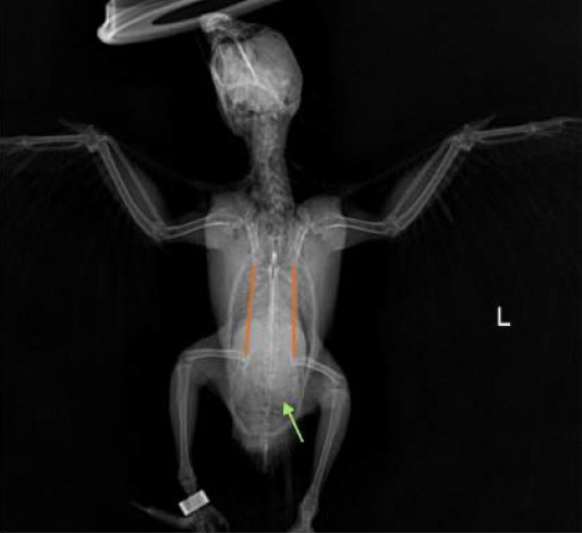 》Figure 3: VD view. The silhouette of liver lobes was obviously extended, indicating the potential hepatomegaly. Secondly, the images of GI tracts were distended, taking up spaces in the coelomic cavity.
|
 |
The results of blood tests showed numerous abnormalities in multiple organs, including the liver and kidneys. The packed cell volume (PCV) percentage was 64%, exceeding the normal range of 42-54%. This result suggested that the patient was badly dehydrated, which could cause artifacts in subsequent biochemistry tests.
A shockingly raised level of gamma-glutamyl transferase (GGT), along with significantly decreased aspartate aminotransferase (AST) and total bile acid levels, indicated severe liver disease. Although a sole elevation in GGT is considered inconsistent and insensitive for liver problems due to its wide presence in hepatocytes, bile duct, and renal tubular epithelium (Braun JP, et al., 1983), the correlation among GGT, AST, and total bile acid indexes collectively pointed towards obvious hepatitis. To rule out any possibility of infection, globulin levels and blood smears were examined. The results showed no signs of inflammation or toxic changes in white blood cells, leading to a more reasonable assumption that the liver problem resulted from a long-term imbalanced diet and lack of essential nutrients.
In addition to hepatic enzymes, urine acid and blood calcium levels also exhibited grave changes. These alterations resulted from a decrease in glomerular filtration rate (GFR), indicating that the patient was facing life-threatening acute renal injury. This situation could be a sequential result of the original hepatitis, which caused lethargy, anorexia, and inadequate water intake.
》Table 3: 2024/01/19 Biochemistry results
| Test item | Result | REF. Range/units |
| Glucose | 154 | 249 - 363 mg/dL |
| TBA | 1 | 44 - 108 μmol/L |
| UA | 28.9 | 3.5 - 11 mg/dL |
| PHOS | 3.2 | 3.2 - 4.8 mg/dL |
| CA | 17 | 7.3 - 10.7 mg/dL |
| TP | 3.0 | 2.4 - 4.8 g/dL |
| ALB | 1.0 | 1 - 1.8 g/dL |
| GLOB | 2.0 | 0 - 9999 g/dL |
| AST | 5 | 160 - 383 U/L |
| GGT | 2000 | 1 - 30 U/L |
| CHOL | 262 | 140 - 360 mg/dL |
| CK | 50 | 58 - 245 U/L |
| LDH | 114 | 120 - 455 U/L |
This case could be underscored as simply a beak abnormality without comprehensive blood tests. The seemingly nonspecific signs were actually potential indicators of serious underlying health conditions in avian patients. A tailored treatment plan focusing on supportive care and anti-inflammatory agents was introduced to the patient. The supportive therapy aimed to maintain both basic nutrient requirements through tube feeding four times a day and ensure proper hydration by administering lactated Ringer’s solution subcutaneously twice daily. Oral medication primarily included silymarin, an antioxidant known for its hepatoprotective properties. Silymarin has been shown to act as a free radical scavenger and modulate enzymes associated with cellular damage, making it a valuable component in managing liver diseases. Clinical studies have demonstrated its effectiveness in reducing liver-related mortality and improving liver function in various conditions, including fatty liver disease and hepatitis. By reducing oxidative stress and protecting liver cells from further damage, silymarin plays a crucial role in the recovery process for patients with hepatic dysfunction.
| It is important to note that while it may seem appropriate to use non-steroidal anti-inflammatory drugs (NSAIDs) for the ongoing hepatitis, their well-known side effect of decreasing renal blood flow could lead to further damage, especially since the patient was already suffering from acute kidney injury (AKI). After being hospitalized for eight days, the conure parrot showed signs of recovery and was subsequently discharged with a plan for outpatient treatment over the next two months. | 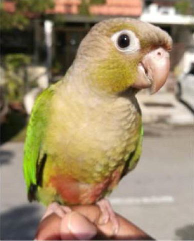 |
Case 3
| An African gray parrot presented with a chief complaint of anorexia and dullness for more than a week. The bird had been taken to two other clinics for oral prescriptions but showed no signs of recovery. According to the owner, the parrot lost its appetite for cereal feedings and was only willing to consume some fruits. | 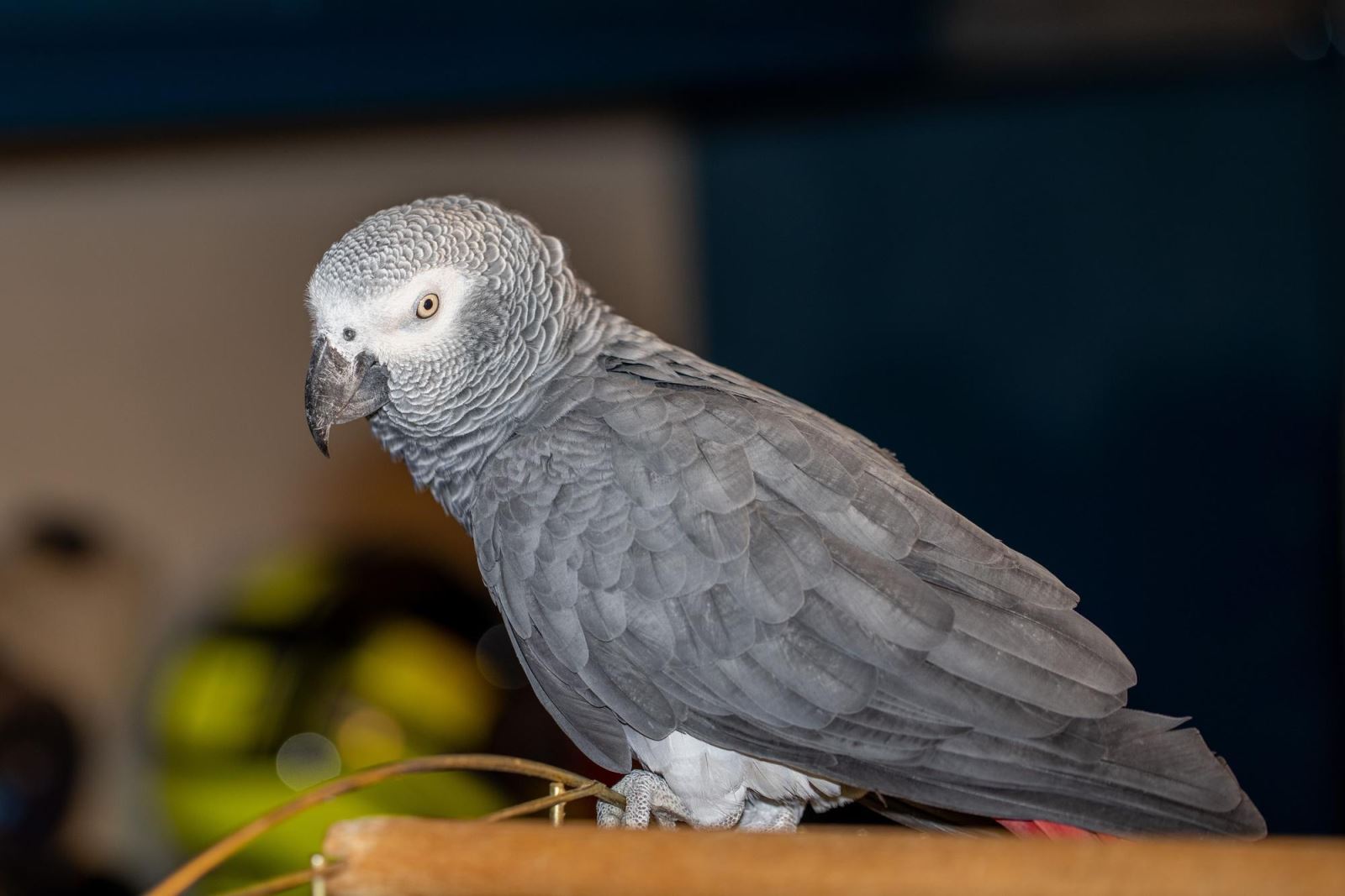 |
Upon examination in the clinic, the parrot displayed a subdued demeanor, slight shivering, half-closed eyes, and sunken eye pits. The stool was dark green and covered in mucus, and a considerable amount of urine was observed, with a notable lack of uric acid. These signs indicated that the patient was very uncomfortable and suffering from more than 5% dehydration. Auscultation revealed unremarkable findings, but palpation indicated slight swelling of the organs within the body cavity. The clinical signs observed in this African gray parrot suggest significant underlying health issues, particularly related to renal function. The combination of anorexia, lethargy, and dehydration points towards a potential renal compromise or systemic illness. The dark green stool covered in mucus may indicate gastrointestinal distress or malabsorption, further complicating the clinical picture. The presence of polyuria alongside dehydration is particularly concerning; it suggests that the kidneys are unable to concentrate urine effectively, which can be indicative of renal disease or other metabolic disorders. The results of the physical examination thus prompted a recommendation for thorough examinations including radiography and blood works. No sedation was needed while the procedure conducted, with the blood sample taken from the medial metatarsal vein.
 |
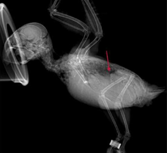
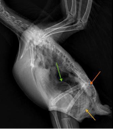 》Figure 6: Right recumbency lateral view. The opacity of kidney was increased, and the size of the silhouette was enlarged. Noted that the proventriculus was dilated with excessive gas accumulation
|
The imaging results indicated that the kidney of the African gray parrot was obviously swollen, with increased opacity, suggesting possible nephritis. Additionally, the walls of the gastrointestinal lumens were clearly thickened, indicating ongoing inflammation. Excessive gas accumulation was noted in the proventriculus, which may signify abnormal gastrointestinal motility and inflammation. Surprisingly, the liver appeared unremarkable in the imaging studies.
The packed cell volume (PCV) of the parrot was measured at 29%, significantly lower than the normal range of 45-53%. Considering the dehydration status, this number would likely be even lower, indicating that the patient was suffering from severe anemia. Renal function was severely compromised, as evidenced by significantly decreased levels of urea and phosphorus. The kidneys had lost their ability to concentrate urine effectively, resulting in an inability to retain these solutes. Other electrolytes, such as sodium and chloride, were also affected due to marked dehydration. Fortunately, potassium levels remained within the normal range, which facilitated easier management of the electrolyte imbalance.
These findings align with established knowledge regarding avian renal disease. Acute renal failure in birds can result from various factors, including ischemic or toxic insults, and is often characterized by a sudden decrease in renal function (Cojean O, et al., 2020). The observed nephritis could be due to infectious agents or other underlying conditions that compromise renal integrity (Schmidt RE. 2006). The thickened gastrointestinal walls and excessive gas accumulation further complicate the clinical picture, suggesting potential gastrointestinal disease that may be contributing to the parrot's overall condition.
The creatinine kinase (CK) level and lactate dehydrogenase (LDH) level were also noted in the results. Combined with elevated globulin levels, it is clear that the patient was suffering from systemic inflammation. Given the radiographic findings of gastrointestinal tract inflammation, it is plausible that this inflammatory situation resulted from dietary factors. Upon questioning the owner, it was revealed that the parrot had been offered salted peanuts as a treat a week prior to symptom onset, coinciding with all observed symptoms.
These insights suggest that dietary indiscretion may have played a significant role in exacerbating the parrot's condition. Salted peanuts could lead to electrolyte imbalances and gastrointestinal distress in birds not accustomed to high salt intake. The combination of systemic inflammation and renal compromise necessitates a comprehensive treatment approach focusing on hydration, nutritional support, and monitoring of renal function.
》Table 4: 2024/11/02 Biochemistry results
| Test item | Result | REF. Range/units |
| Glucose | 266 | 206 - 275 mg/dL |
| TBA | 1 | 12 - 96 μmol/L |
| UA | 1.5 | 2.7 - 8.8 mg/dL |
| PHOS | 2.0 | 3.2 - 5.4 mg/dL |
| CA | 9.2 | 7.7 - 11.3 mg/dL |
| TP | 4.7 | 3.2 - 5.2 g/dL |
| ALB | 2.2 | 1.22 - 2.52 g/dL |
| GLOB | 2.5 | 0.4 - 1.9 g/dL |
| AST | 296 | 109 - 305 U/L |
| GGT | 3 | 1 - 10 U/L |
| CHOL | 218 | 160 - 425 mg/dL |
| CK | 748 | 228 - 322 U/L |
| LDH | 576 | 145 - 465 U/L |
| Sodium | 157 | 157 - 165 mEq/L |
| Patassium | 3.4 | 2.9 - 4.6 mEq/L |
| Chloride | 147 | 115 - 125 mEq/L |
The treatment plan for the African gray parrot focused on fluid therapy to alleviate the ongoing acute renal failure, administering Lactated Ringer's solution subcutaneously at a dosage of 15 mL twice daily. Anti-inflammatory agents, specifically meloxicam, were used cautiously alongside sufficient fluid support, with famotidine included to protect the gastrointestinal mucosa. Nutritional support was also a critical component of the treatment; gavage feeding was conducted, providing 10 mL of a liquid diet supplemented with vitamins four times a day.
Remarkably, the patient quickly regained its appetite by the second day of hospitalization and began exhibiting feather preening behavior. These positive responses to treatment led to an early discharge after three days of intensive care, with the parrot sent home with prescribed oral medications.

Conclusion
The nutritional needs of psittacine birds, particularly common pet species, are diverse and often misunderstood by their owners. While species such as budgerigars, cockatiels, and hyacinth macaws are primarily granivorous, frugivorous species like Amazon parrots and most macaws, nectarivorous lorikeets, and omnivorous African gray parrots all require varied diets to thrive (Hess L. et al., 2002). Unfortunately, many pet owners default to cereal-based diets for their birds due to convenience, which can lead to significant nutritional deficiencies. Even granivorous species that naturally consume a wide variety of seeds in the wild may struggle to meet their nutritional requirements when offered a limited diet.
Research indicates that healthy parrots require lower levels of fat, protein, and calcium compared to poultry species; however, the common cereal-based products often provide excessive calories. This caloric surplus can lead to decreased motivation to eat a varied diet, resulting in malnutrition. Studies have shown that cereal diets frequently lack essential nutrients such as amino acids (methionine and lysine), vital minerals (calcium and iodine), and important vitamins (A and D) (Hess L. 2020).
To ensure optimal health and well-being for pet birds, it is crucial for owners to understand the specific dietary needs of their avian companions. A balanced diet should include a variety of fresh fruits, vegetables, and high-quality pellets designed specifically for psittacines. These formulated diets can help prevent deficiencies commonly associated with seed-based feeding practices. Furthermore, educating pet owners about the importance of dietary diversity can lead to healthier birds with improved overall health outcomes.
References:
This case study was conducted by Chung-Jui Chen, Director of Jungle Exotic Animal Hospital, and Zheng-You Zhuang, DVM. The biochemical profile tests in cases here were measured by AmiShield veterinary chemistry analyzer and the exclusive Avian/Reptile Profile Panels.
1. Bavelaar, F.J. & Beynen, Anton. "Influence of amount and type of dietary fat on plasma cholesterol concentrations in African grey parrots." J Appl Res Vet Med. 1. 1-8, 2003.
2. Bandyopadhyay, Samiran. "Systemic Clinical and Metabolic Diseases." Pet bird diseases and care 167–252. 25 Feb. 2017, doi:10.1007/978-981-10-3674-3_3.
3. Braun JP, Benard P, Burgat V, Rico AG. "Gamma glutamyl transferase in domestic animals." Veterinary Research Communications 6.1 (1983): 77-90.
4. Cojean O, Larrat S, Vergneau-Grosset C. Clinical Management of Avian Renal Disease. Vet Clin North Am Exot Anim Pract. 2020;23(1):75-101. doi:10.1016/j.cvex.2019.08.004.
5. Elliot, Dorianne. "Recognising Avian Malnutrition." World Small Animal Veterinary Association World Congress Proceedings, 2014.
6. Harr, K. E. "Clinical Chemistry of Companion Avian Species: A Review. Veterinary Clinical Pathology", 31(3), 141–142. doi:10.1111/j.1939-165x.2002.tb00295.x, 2002.
7. Hess, L., Mauldin, G., & Rosenthal, K. (2002). Estimated nutrient content of diets commonly fed to pet birds. Veterinary record, 150(13), 399-404.
8. Hess, L. (2020). Companion Parrot Nutrition—Nutrient Requirements, Common Feeding Practices, and Nutrition-Related Diseases ExoticsCon Virtual 2020 Proceedings.
9. Schmidt RE. Types of renal disease in avian species. Vet Clin North Am Exot Anim Pract. 2006; 9(1): 97-106. doi:10.1016/j.cvex.2005.10.003.
Read more avian case studies:
Avian Bornavirus in Parrots