


2024
10.25
Introduction
In recent years, the part of rabbits seen in small animal clinics has been increasing annually. Rabbits are timid and shy animals that often hide their illnesses, so owners usually feel some wrong in their pet, but they don't know the reason why. However radiology diagnosis and physical examinations results often not enough accurate. Hematological examination may help us better understand the animal's condition, and unexpected findings are often discovered in the results.
  |
Case 1
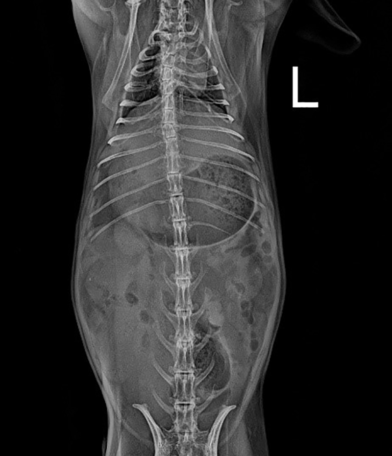
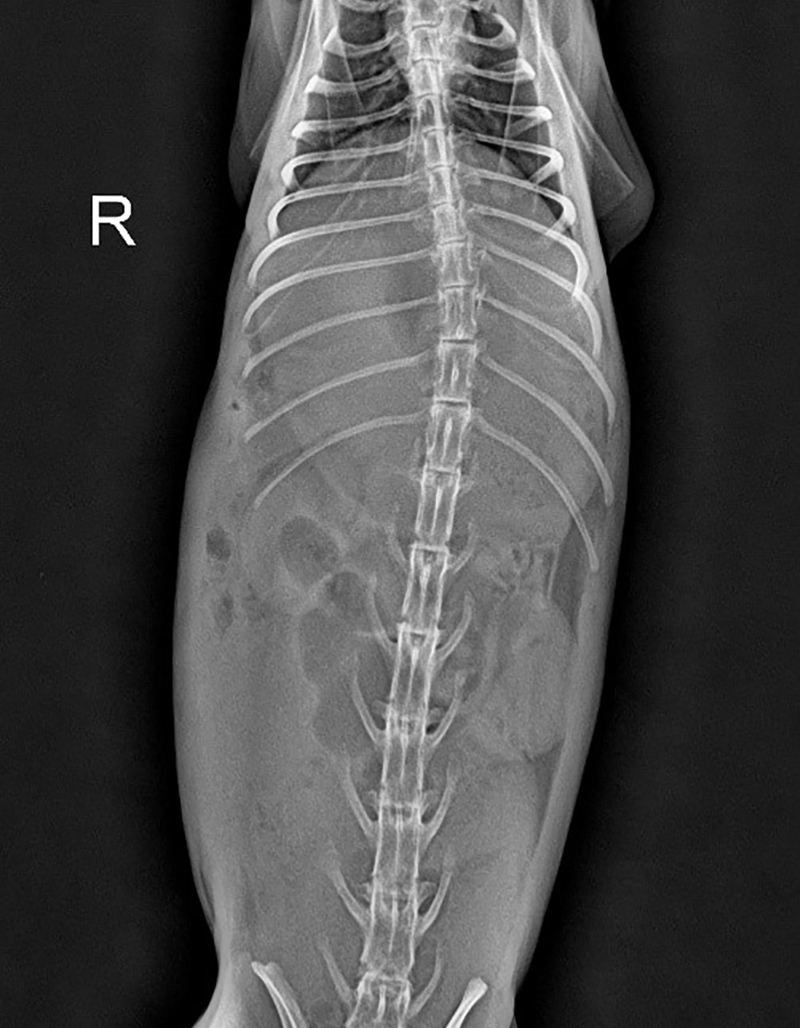
| This is a 6-year-old spayed female rabbit with a history of gastric obstruction and nasolacrimal duct blockage. Once a week, jujube fruit and goji berries are given as snacks. The owner has noticed a decrease in the rabbit's energy, appetite, and activity levels for one day. Imaging diagnostics revealed significant gas accumulation in the stomach and a small amount of gas accumulation in the cecum (shown as Figure 1). |
 |
 |
 |
|
| A large amount of gas is visible in the stomach | ||
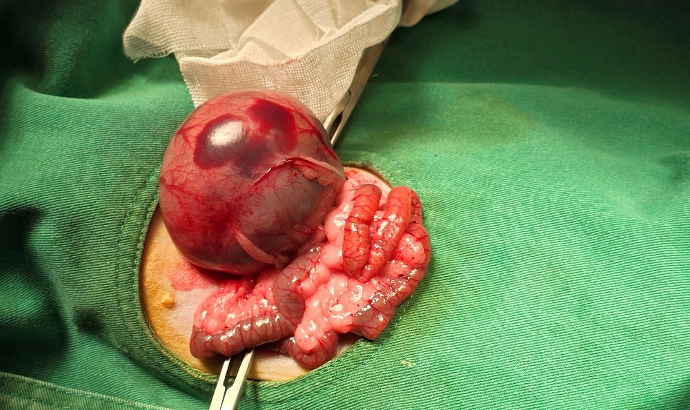
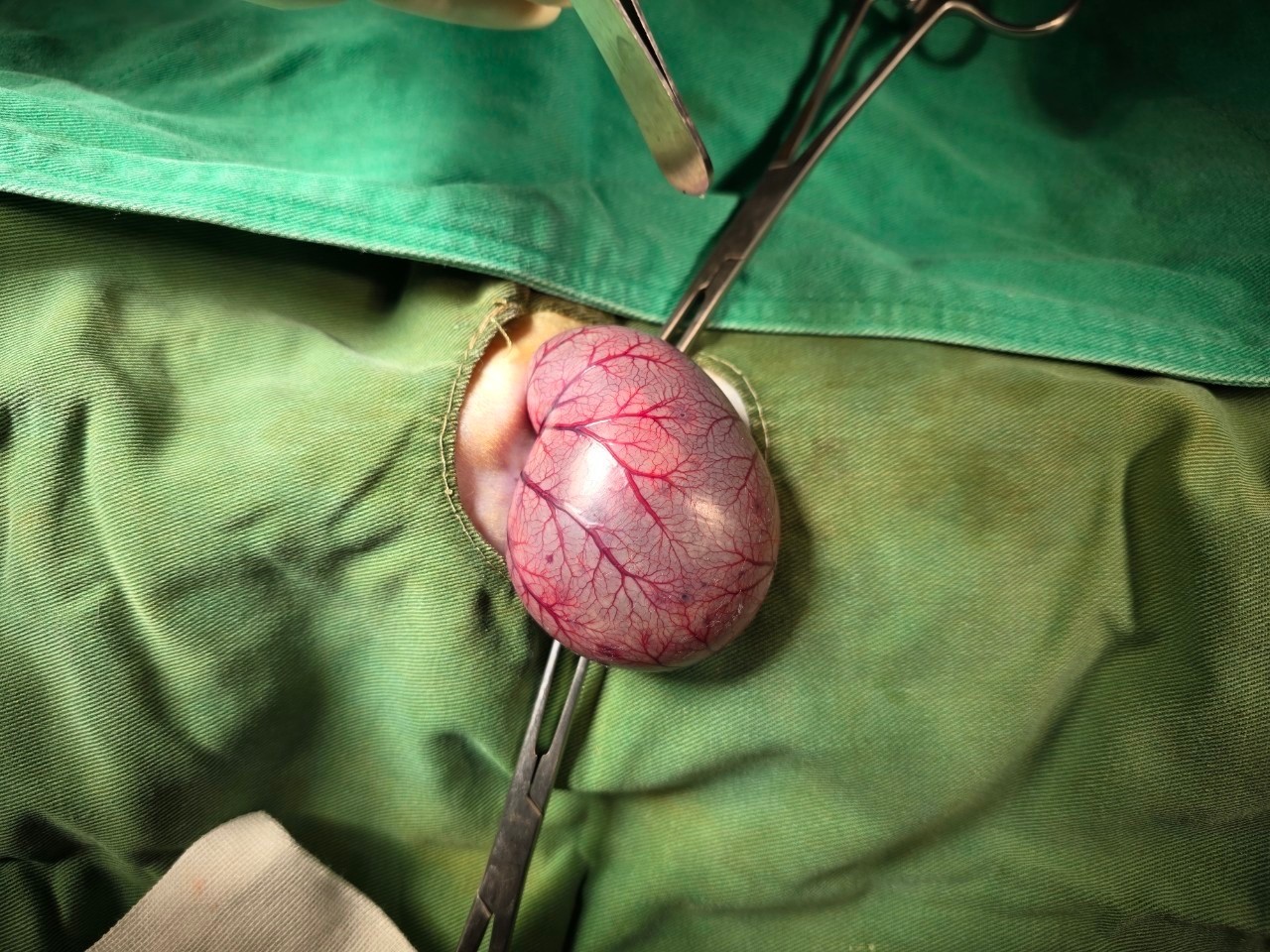
After discussing with the owner, initial medical treatment was administered, including analgesics and intravenous fluid therapy. However, on 09/03, the animal's symptoms showed no improvement. There was no observation of defecation, and X-rays indicated that the stomach remained significantly distended. A high likelihood of gastric obstruction was suspected. Following further discussion with the owner, surgical intervention was performed on 09/03. During the surgery, it was discovered that there was a foreign body in the duodenum causing intestinal perforation (shown as Figure 2).
 |
 |
| Diffuse redness is visible on the serosal surface of the gastrointestinal tract | Location of foreign body perforation |
On the second postoperative day, 09/05, a hematological follow-up was conducted, revealing that the animal's biochemical indices were elevated beyond normal ranges (shown as Table 1). Additionally, the animal continued to exhibit lethargy and poor appetite, and there was no defecation observed post-surgery. After evaluation, the prognosis for the animal was deemed poor, leading to the final decision to proceed with euthanasia.
》Table 1: Biochemistry results
| Test item | 09/05 | 09/02 | REF.R Range/units |
| Glucose | 152 | 193 | 75-150 mg/dL |
| AMY | 378 | 148 | 50-300 U/L |
| CREA | 5.9 | 1.6 | 0.3-2.5 mg/dL |
| BUN | 97 | 52 | 13-29 mg/dL |
| PHOS | 10.2 | 2.8 | 2.3-6.9 mg/dL |
| CA | 10.7 | 9.4 | 5.6-14.8 mg/dL |
| TP | 6.8 | 6.9 | 5.4-7.5 g/dL |
| ALB | 2.5 | 2.8 | 2.5-5 g/dL |
| GLOB | 4.3 | 5.6 | 1.5-3.5 g/dL |
| ALB / GLOB | 0.6 | 2.0 | 0-9999 |
| ALT | 237 | 50 | 14-80 U/L |
| AST | 474 | 29 | 14-113 U/L |
| ALP | 29 | 44 | 70-145 U/L |
| GGT | 5 | 9 | 1-15 U/L |
| TBIL | 0.15 | 0.7 | 0.1-0.8 mg/dL |
Case 2
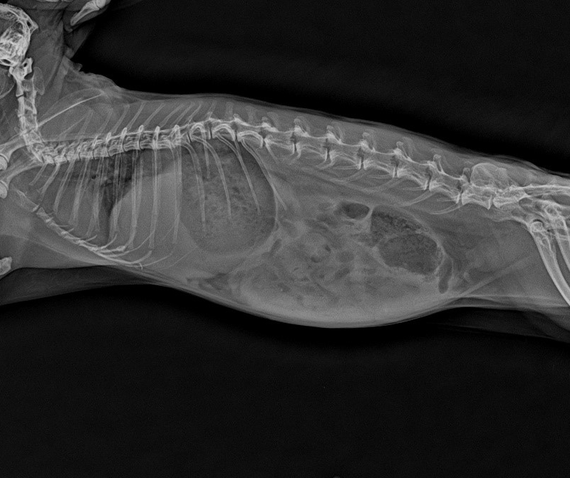
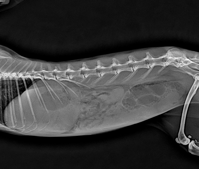
| This is a 16-month-old intact male rabbit with congenital left stifle (knee) dysplasia. The owner reports decreased appetite and mild diarrhea for the past two days, prompting the veterinary visit. Imaging studies revealed a "fried egg" appearance in the stomach, highly suggestive of acute gastric dilatation. Biochemistry results showed markedly elevated blood glucose levels. Based on the clinical signs and biochemistry test results, the veterinarian strongly suspected the rabbit was suffering from severe pain due to gastric dilatation. Emergency surgical intervention was immediately performed to address the condition. |
 |
》Table 2: Biochemistry results
| Test item | 03/27 | 03/18 | 03/14 | REF.R Range/units |
| Glucose | 147 | 147 | 405 | 75-150 mg/dL |
| AMY | 88 | 159 | 757 | 50-300 U/L |
| CREA | 0.9 | 0.8 | 1.5 | 0.3-2.5 mg/dL |
| BUN | 18 | 12 | 114 | 13-29 mg/dL |
| PHOS | 3.6 | 2.1 | 12.1 | 2.3-6.9 mg/dL |
| CA | 12.8 | 11.5 | 12.3 | 5.6-14.8 mg/dL |
| TP | 6.0 | 5.7 | 7.9 | 5.4-7.5 g/dL |
| ALB | 2.9 | 2.7 | 2.6 | 2.5-5 g/dL |
| GLOB | 3.1 | 3.0 | 5.3 | 1.5-3.5 g/dL |
| ALB / GLOB | 0.9 | 0.9 | 0.5 | 0-9999 |
| ALT | 76 | 199 | 43 | 14-80 U/L |
| AST | 18 | 79 | 44 | 14-113 U/L |
| ALP | 203 | 210 | 205 | 70-145 U/L |
| GGT | 14 | 14 | 14 | 1-15 U/L |
| TBIL | 0.57 | 0.33 | 0.2 | 0.1-0.8 mg/dL |
 |
 |
|
| Imaging studies revealed a "fried egg" appearance in the stomach | ||
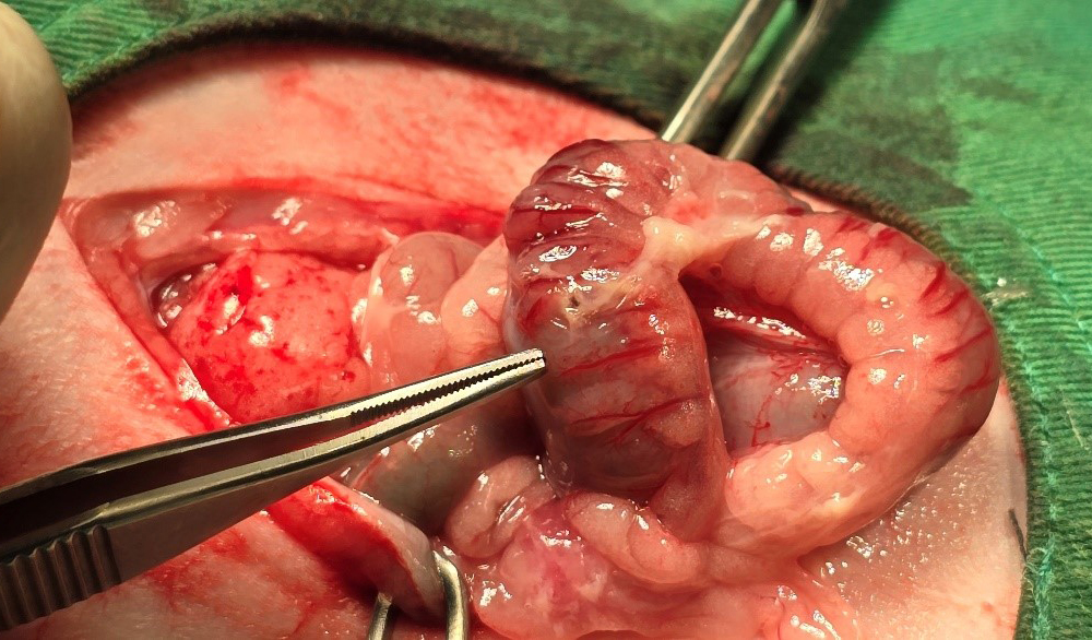
Due to the rabbit's congenital stifle (knee) joint dysplasia, there is chronic joint wear and tear, leading to persistently elevated alkaline phosphatase (ALP) levels outside the normal range. On the biochemistry test performed on 03/14, the rabbit was found to have severely elevated blood glucose levels. Imaging studies also revealed a "fried egg" appearance in the stomach, diagnostic for acute gastric dilatation. After discussion with the owner, surgical intervention was elected to address the suspected gastric obstruction.
 |
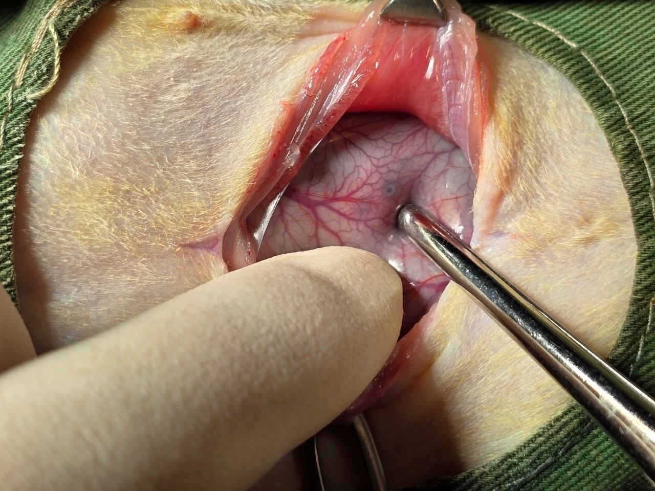 |
| The serosal surface of the stomach shows vascular congestion, and suspected multiple localized ulcerations are visible | Suspected ulceration sites |
On recheck biochemistry test 4 days after surgery, the rabbit was found to have elevated alanine aminotransferase (ALT) levels, likely due to free radical reperfusion injury and potential refeeding syndrome following the procedure. The amylase (AMY) level was elevated on 03/18, potentially indicating pancreatic injury, which can be caused by conditions such as pancreatitis, peritonitis, abdominal trauma, or external compression. At the recheck examination on 03/28 after discharge, the only abnormality remaining was the persistently elevated ALP, while all other parameters had normalized.
Conclusion
Rabbits are prey species that tend to mask their clinical signs of illness, often leaving owners unaware of an underlying disease process. Owners typically only recognize nonspecific symptoms such as lethargy, inappetence, and abnormal defecation before seeking veterinary care. In herbivores, gastrointestinal disturbances can rapidly become life-threatening.
When the owner is unable to provide detailed historical information, performing comprehensive bloodwork can be a valuable diagnostic tool to better understand the rabbit's physiologic status and facilitate an accurate diagnosis. Rabbits' ability to conceal their disease states underscores the importance of proactive preventive care and vigilant monitoring by owners to identify subtle changes that may indicate an emerging health concern.
References:
This case study was conducted by Chung-Jui Chen, Director of Jungle Exotic Animal Hospital, and Yong-Chen Zheng, DVM. The biochemical profile tests in cases here were measured by AmiShield veterinary chemistry analyzer and the exclusive Comprehensive Plus Panels.
1. Harcourt-Brown F.M., Baker S.J. Parathyroid hormone, hematological and biochemical parameters in relation to dental disease and husbandry in pet rabbits. J Small Anim Pract. 2001;42:130–136.
2. Fudge A.M. Rabbit hematology. In: Fudge A.M., editor. Laboratory Medicine: Avian and Exotic Pets. WB Saunders Company; Philadelphia, PA: 2000. pp. 273–275.
3. Saunders R.A., Davies R.R. Blackwell Publishing; Oxford, UK: 2005. Notes on Rabbit Internal Medicine.
4. Hayashi, H., Imanishi, N., Ohnishi, M. and Tojo, S.J. 2001. Nephron 87: 352-360.
5. Meredith, A., & Lord, B. (Eds.). (2014). BSAVA manual of rabbit medicine. British Small Animal Veterinary Association.
6. Carpenter, J. W., & Harms, C. A. (Eds.). (2023). Exotic animal formulary (6th ed.). Wiley Blackwell.