


2024
07.03
Acute phase proteins (APPs) are gaining prominence in veterinary medicine for their role in diagnosis, prognosis and treatment monitoring. Despite extensive research and the availability of specific assays, their application in veterinary practice is still limited compared to human medicine.
APPs are liver-synthesized proteins responding to pro-inflammatory cytokines during the acute phase reaction (APR). They show various responses to pathologic conditions, such as infections and tumors. APPs are more effective than WBC counts in detecting early inflammation and for identifying early remission or recurrence of diseases. They are valuable in determining the appropriate duration of antimicrobial treatments. Measuring APPs is particularly useful in cats with feline infectious peritonitis (FIP) to help differentiate between those with FIP and those with similar symptoms but without the disease.
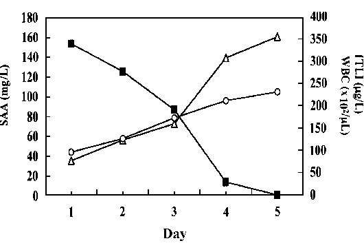
》Figure 1: Time–course of changes in SAA concentration (■), WBC count (O), and fTLI concentration (△) in a cat presenting with pancreatitis on day 1 and during 5 days of treatment with plasma transfusion, intravenous fluids, prednisolone, and antibiotics.
》Table 1: According to the increase in serum concentration of APPs during acute reactions, they can be divided into three types: major, moderate and minor.
| APP Classification | Serum Concentration Change | Reaction Time | Examples |
| Major APP | Increase >1000-fold | Peak within 24-48hr |
Serum amyloid A (SAA)
|
| Moderate APP | Increase 5- to 10-fold | Peak after 2-3 days |
alpha-1-acid glycoprotein (AGP), haptoglobin
|
| Minor APP | Increase max. 2-fold | N/A |
C-reactive protein, ceruloplasmin
|
SAA and AGP are the most extensively investigated APPs in cats. The investigations in different diseases are listed in Table 3. Examples of their kinetic responses in cats subjected to ovariohysterectomy and gastrotomy are shown below. (For more SAA Clinical Application, please refer to "Case Study : fSAA Clinical Application")
》Table 2: Comparison of SAA and AGP
| APP Type | Reaction Time | Maintenance | Magnitude of Increase |
| SAA | Both peaked in 1-2 days |
Less than 5 days
|
Greater (27-fold) – in proportion to severity of inflammation
|
| AGP |
For weeks
|
Smaller (4-fold)
|
Elevated SAA levels are observed in variable inflammatory diseases. However, APPs cannot distinguish between septic and nonseptic inflammation. APPs also have limited specificity for different pathologic conditions, while FIP is a notable exception. Serum AGP levels >1.5 g/L or AGP levels in abdominal effusion >1550 ug/ml provide high sensitivity and specificity in FIP diagnosis.
Moreover, sequential APP measurements can track disease progression and therapeutic responses. For example, SAA levels increase at the onset or reoccurrence of feline pancreatic lipase immunoreactivity, and decline as it resolves. AGP levels track treatment response of feline chronic gingivostomatitis. However, the prognostic properties of APPs remain arguable, and this may be improved with the combined measurement of multiple APPs.
Future applications of APPs in veterinary medicine may mirror human medicine, including animal welfare assessment, guidance of antibiotic treatment, the use of an APP profile for sepsis screens and acute phase index for inflammation detection. Development of new inflammatory markers is also promising. Paraoxonase-1 (PON-1) acts as a negative acute APP in cats associated with lipid oxidation, while procalcitonin (PCT) has the potential to distinguish between infectious and noninfectious inflammation.
In conclusion, APPs are valuable diagnostic tools for cats with systemic inflammation, especially FIP. Rigorous assay validation and standardization are crucial for their broader application. Continued research and the development of new markers and POC assays will likely expand their clinical utility.
》Table 3: Positive acute phase proteins investigated in different diseases in cats
| Disease | Acute Phase Protein | Magnitude of Increase | References |
| FIP | AGP Haptoglobin SAA |
17x 10x 2x 144x |
Paltrinieri et al2 Duthie et al3 Duthie et al3 Yuki et al4 Tecles et al5 |
| Congestive heart failure | SAA | Not specified | Liu et al6 |
| Upper respiratory tract infections
Pneumonia
Pancreatitis |
SAA |
140x 134x 4x |
Yuki et al4 |
| Sepsis | SAA | 43x | Troia et al7 |
| Pyometra |
SAA Haptoglobin |
155x 167x 2x |
Yuki et al8 Vilhena et al9 |
| Hyperthyroidism |
SAA AGP Haptoglobin |
Not specified Not specified Not specified |
Glück et al10 |
| CKD | SAA | 2x | Javard et al11 |
| Dirofilaria immitis and Wolbachia (with clinical signs) | SAA Haptoglobin |
100x 2x |
Silvestre-Ferreira et al12 |
| Hepatozoon felis Babesia vogeli |
SAA Haptoglobin Haptoglobin |
169x 8x 4x |
Vilhena et al13 |
| Gingivostomatitis | AGP SAA |
1-3x 4x |
Mestrinho et al14 Yuki et al8 |
| Injury
Renal failure
Infectious diseases
FLUTD Diabetes mellitus |
SAA |
102x 52x 78.7x 47x 13x |
Sasaki et al15 |
| Various tumors grouped in macro-categories: carcinoma, sarcoma, and discrete round cell tumors (lymphoma, mast cell tumor, and melanoma) in Sasaki et al study Nonspecified tumors in Selting et al study |
SAA AGP |
28x 2x |
Sasaki et al15 Selting et al16 |
| Lymphoma | AGP SAA Haptoglobin |
2x 3x 10x 2x |
Correa et al17 Winkel et al18 Winkel et al18 Love et al19 |
| IBD | Haptoglobin | 2x | Love et al19 |
Note: This figure is adapted from "Acute phase proteins in cats: Diagnostic and prognostic role, future directions, and analytical challenges", by Gabriele Rossi, 2023, Vet Clin Pathol. 2023;52(Suppl. 1), P.39.
Reference:
Special thanks to Ellie Hsiao-Mei Chang, a student at National Chunghsing University, for her help in organizing the abstracts of literature.
1. Rossi G. Acute phase proteins in cats: Diagnostic and prognostic role, future directions, and analytical challenges. Vet Clin Pathol. 2023;52(Suppl. 1):37-49. doi:10.1111/vcp.13238
2. Paltrinieri S, Giordano A, Tranquillo V, Guazzetti S. Critical assessment of the diagnostic value of feline alpha1-acid glycoprotein for feline infectious peritonitis using the likelihood ratios
3. Duthie S, Eckersall PD, Addie DD. Value of alpha-1 acid glycoprotein in the diagnosis of feline infectious peritonitis. Vet Rec. 1997;141:299-303.
4. Yuki M, Aoyama R, Nakagawa M, Hirano T, Naitoh E, Kainuma D. A clinical investigation on serum amyloid a concentration in client-owned healthy and diseased cats in a primary care animal hospital. Vet Sci. 2020;7(2):45. doi:10.3390/vetsci7020045
5. Tecles F, Caldín M, Tvarijonaviciute A, Escribano D, Martínez-Subiela S, Cerón JJ. Serum biomarkers of oxidative stress in cats with feline infectious peritonitis. Res vet Sci. 2015;100:12-17. doi:10.1016/j.rvsc.2015.02.007
6. Liu M, Köster LS, Fosgate GT, et al. Cardiovascular-renal axis disorder and acute-phase proteins in cats with congestive heart failure caused by primary cardiomyopathy. J vet Intern Med. 2020;34(3):1078-1090. doi:10.1111/jvim.15757
7. Troia R, Gruarin M, Foglia A, Agnoli C, Dondi F, Giunti M. Serum amyloid a in the diagnosis of feline sepsis. J vet Diagn Invest. 2017;29(6):856-859. doi:10.1177/1040638717722815
8. Zachary J. Inflammation and healing. Pathologic basis of veterinary disease. 6th ed. Elsevier; 2017:73-131.
9. Vilhena H, Figueiredo M, Ceron JJ, et al. Acute phase proteins and antioxidant responses in queens with pyometra. Theriogenology. 2018;115:30-37. doi:10.1016/j.theriogenology.2018.04.010
10. Glück K, Mohrs S, Hazuchova K, Bauer N, Neiger R. Impact of radioiodine treatment on acute phase proteins in hyperthyroid cats. J Feline Med Surg. 2022;24(4):359-365. doi:10.1177/1098612X211024954
11. Javard R, Grimes C, Bau-Gaudreault L, Dunn M. Acute-phase Proteins and iron status in cats with chronic Kidney disease. J vet Intern Med. 2017;31(2):457-464. doi:10.1111/jvim.14661
12. Silvestre-Ferreira AC, Vieira L, Vilhena H, et al. Serum acute phase proteins in Dirofilaria immitis and Wolbachia seropositive cats. J Feline Med Surg. 2017;19(6):693-696. doi:10.1177/1098612X15625435
13. Vilhena H, Tvarijonaviciute A, Cerón JJ, Vieira L, Pastor J, Silvestre-Ferreira AC. Acute phase proteins response in cats naturally infected with Hepatozoon felis and Babesia vogeli. Vet Clin Pathol. 2017;46(1):72-76. doi:10.1111/vcp.12451
14. Mestrinho LA, Rosa R, Ramalho P, et al. A pilot study to evaluate the serum Alpha-1 acid glycoprotein response in cats suffering from feline chronic gingivostomatitis. BMC vet Res. 2020;16(1):390. doi:10.1186/s12917-020-02590-2
15. Sasaki K, Ma Z, Khatlani S. Evaluation of feline serum amyloid a (SAA) as an inflammatory marker. J vet Med Sci. 2003;65:545-548
16. Selting KA, Ogilvie GK, Lana SE. Serum alpha 1-acid glycoprotein concentrations in healthy and tumor-bearing cats. J vet Intern Med. 2000;14:503-506
17. Correa SS, Mauldin GN, Mauldin GE. Serum alpha 1-acid glycoprotein concentration in cats with lymphoma. J Am Anim Hosp Assoc. 2001;37:153-158.
18. Winkel VM, Pavan TL, Wirthl VA, Alves AL, Lucas SR. Serum alpha-1 acid glycoprotein and serum amyloid a concentrations in cats receiving antineoplastic treatment for lymphoma. Am J vet Res. 2015;76(11):983-988. doi:10.2460/ajvr.76.11.983
19. Love EK, Leibman NF, Ringold R, Lamb K. Serum haptoglobin concentrations in feline inflammatory bowel disease and small-cell alimentary lymphoma: a potential biomarker for feline chronic enteropathies. J Feline Med Surg. 2021;23(10):959-964. doi:10.1177/1098612X21991448
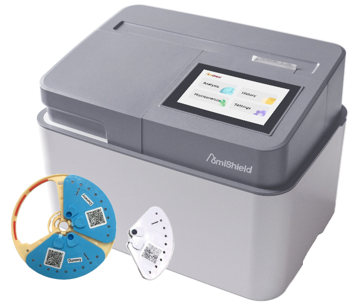
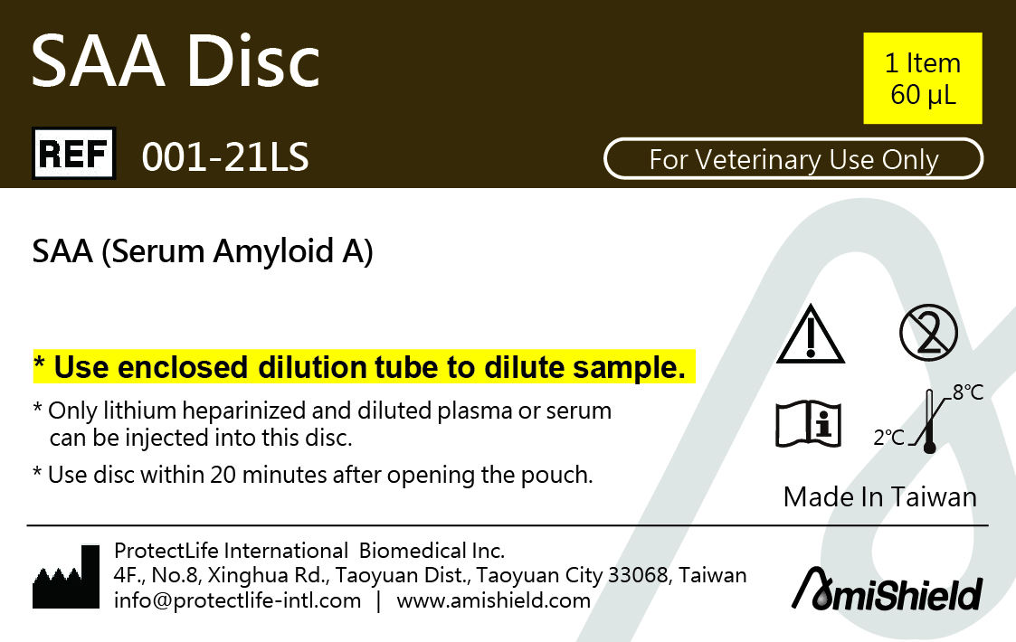
AmiShield SAA DiscThe AmiShield SAA disc can quantitatively detect SAA in lithium heparinized plasma or serum to assist the veterinarian in diagnosing Infectious, inflammatory diseases, tissue injury, malignant tumors, tissue necrosis, and surgery.AmiShield CRP DiscThe AmiShield CRP disc can quantitatively detect CRP in lithium heparinized whole blood, plasma or serum to assist the veterinarian in diagnosing Infectious, inflammatory diseases, tissue injury, inflammatory bowel disease, allergic, and immune mediated disease.Here is the reference range table:
|
   |